
 Our Centers
 Select City


 Select City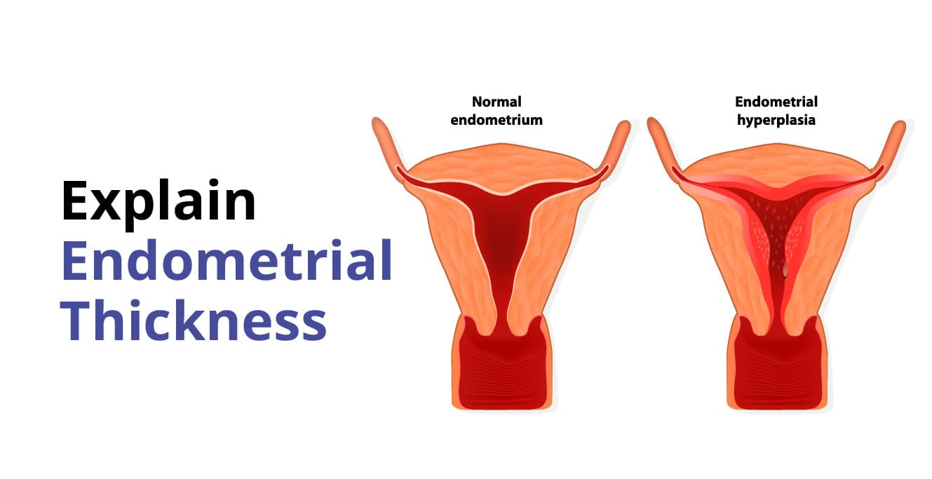
What is Endometrial Thickness? How is it Measured?

Table of Contents
- What is Endometrial Thickness?
- What is Considered Normal Endometrial Thickness and How is It Measured?
- Normal Endometrial Thickness Chart: Day-by-Day & Cycle Phase Guide
- Ultrasound Report Decoder: What Do “Endometrial Echoes” and “Trilaminar” Mean?
- What Does Your Exact Number Mean? (Diagnostic Reference Key)
- What Is an Ideal Endometrial Thickness for IVF?
- What is a Thin Endometrium?
- What is a Thickened Endometrium?
- Causes of a Thickened Endometrium
- Symptoms of a Thickened Endometrium
- Diagnosis of a Thickened Endometrium
- Treatment of a Thickened Endometrium
- How to Reduce Endometrial Thickness Naturally (Lifestyle & Hormonal Balance)
- Endometrial Thickness and Cancer: When Should You Worry?
- Conclusion
- Frequently Asked Questions (FAQs)
- Is a 10 mm endometrial thickness normal for pregnancy?
- What is the normal endometrial thickness on Day 10, Day 11, and Day 12 of a cycle?
- Is a 20 mm endometrial thickness dangerous?
- What does “normal endometrial echo” mean on an ultrasound report?
- What does a 15 mm endometrial thickness mean?
- How does a thin endometrium affect IVF success?
- Can diet alone fix a thickened endometrium?
Female reproductive health depends on many factors, and the thickness of the endometrium is one of them. The endometrium is the lining inside the uterus that changes with each menstrual cycle. During the cycle, this lining grows under the influence of hormones so that it is ready to support a fertilised embryo. For this to happen smoothly, the lining needs to reach a suitable thickness. If it remains too thin or becomes thicker than expected, it can affect periods, fertility, and overall reproductive health.
What is Endometrial Thickness?
The endometrium is the inner mucosal lining of the uterus. Endometrial thickness simply refers to how thick this lining is at a given time in your menstrual cycle. Every month, this lining grows thicker in preparation for a possible pregnancy. If pregnancy does not happen, it sheds during your period. If the endometrial lining is too thin or too thick, it can impact the process of embryo implantation.
What is Considered Normal Endometrial Thickness and How is It Measured?
The normal endometrial thickness varies significantly across the menstrual cycle, typically ranging from 2 to 4 mm right after your period to 8 to 14 mm during the periovulatory (implantation) phase in reproductive-age women.
Measurement of Endometrial Thickness (Ultrasound Scan)
Normally, a transvaginal ultrasound (TVS) is used to measure the thickness of the endometrium. This is a quick and safe procedure where a small, lubricated probe is placed inside the vagina to get a high-resolution image of the uterus.
During the scan, the sonologist or fertility specialist looks at the uterus in the midsagittal plane and measures the double-layer thickness from one echogenic outer border to the other across the endometrial cavity near the fundus. The measurement is recorded in millimetres (mm).
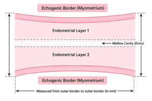
Here is what typically happens during the process:
- You lie down comfortably with an empty bladder while the scan is performed.
- The transvaginal probe provides a detailed view of the uterine cavity.
- The doctor identifies the endometrial lining and measures its thickest point (excluding any fluid within the cavity).
- The timing of the scan matters: For fertility treatments, it is often checked around ovulation or before an embryo transfer, when the lining is expected to reach optimal receptivity.
Normal Endometrial Thickness Chart: Day-by-Day & Cycle Phase Guide
Because hormones change daily, what is “normal” depends on the exact day of your cycle. During an active reproductive cycle, the endometrium typically grows 1 to 2 mm every 24 hours in response to rising estrogen levels:
| Menstrual Phase & Timing | Cycle Days | Typical Thickness (mm) | Ultrasound Pattern & Appearance |
| Menstrual Phase (Early) | Days 1 – 3 | 1 – 3 mm | Thin, broken echogenic line as the lining sheds |
| Post-Menstrual (Baseline) | Days 4 – 5 | 2 – 4 mm | Thin, regular single echogenic line |
| Early Proliferative Phase | Days 6 – 9 | 4 – 7 mm | Hypoechoic (darker) layer with clear outer borders |
| Mid-to-Late Follicular Phase | Days 10 – 11 | 7 – 10 mm | Developing Trilaminar (Triple-Line) pattern |
| Periovulatory (Peak Fertile) | Days 12 – 14 | 8 – 12 mm | Classic Grade A Trilaminar Pattern (Optimal for conception) |
| Secretory (Luteal Phase) | Days 15 – 28 | 10 – 16 mm | Homogeneous, bright hyperechoic (dense) lining |
| Early Pregnancy | Weeks 4 – 6 | 10 – 18 mm | Thick, highly vascular decidualized endometrium |
| Postmenopause (No Bleeding) | Post-Menopause | ≤≤ 8 – 11 mm | Thin, inactive, atrophic lining |
| Postmenopause (With Bleeding) | Post-Menopause | ≤≤ 4 – 5 mm | Strict cutoff; >4 mm>4 mm warrants an endometrial biopsy |
Ultrasound Report Decoder: What Do “Endometrial Echoes” and “Trilaminar” Mean?
When reviewing a pelvic ultrasound or sonography report, you may encounter specific medical terms:
- Endometrial Echo / Echo Complex: Refers to the ultrasound reflection produced by the endometrial stripe. A “normal endometrial echo” means the borders of the lining are smooth, uniform, and well-defined without irregular intracavitary masses.
- Trilaminar (Triple-Line) Pattern: A distinct ultrasound appearance seen right before ovulation consisting of three parallel echogenic lines separated by two dark (hypoechoic) zones. This triple-line appearance is a strong biomarker of a healthy, estrogen-primed lining receptive to embryo implantation.
- Hyperechoic vs. Hypoechoic: Hyperechoic areas appear brighter/white on ultrasound (normal during the secretory phase after ovulation due to mucus and glycogen storage), while hypoechoic areas appear darker (normal during the follicular growing phase).
- Subendometrial Halo: The subtle dark junctional zone separating the endometrium from the myometrium (uterine muscle wall).
What Does Your Exact Number Mean? (Diagnostic Reference Key)
If you recently had a scan and are looking at your measurement, here is what specific thicknesses typically indicate:
1. Less than 6 mm (Thin Endometrium)
- When it is normal: During menstruation (Days 1–5) and after menopause.
- When it is a concern: On Days 11–14 of an ovulation cycle or before an embryo transfer. A lining under 6 mm during the fertile window may reduce implantation rates and requires hormonal or blood-flow support.
2. 7 mm to 8 mm (Borderline to Adequate)
- Considered the standard minimum cutoff for IUI and IVF transfers. If accompanied by a healthy trilaminar pattern, pregnancy success rates remain good.
3. 8 mm to 12 mm (The Ideal Fertile Window)
- The gold standard for natural conception and IVF. Indicates optimal estrogen response, healthy blood flow, and a receptive environment for blastocyst attachment.
- Is 10 mm normal? Yes—a 10 mm trilaminar endometrium around Days 11–14 is considered ideal for embryo implantation.
4. 13 mm to 16 mm (Late Secretory or Early Pregnancy)
- Completely normal in the days leading up to your period (Days 20–28) as progesterone enriches the endometrium, or during the first few weeks of pregnancy.
5. 17 mm to 20 mm or More (Thickened Endometrium)
- Is 20 mm normal? A 20 mm endometrium is thicker than expected.
- In reproductive-age women, it is often caused by anovulatory cycles, PCOS, obesity (excess estrogen), or benign polyps.
- In postmenopausal women, an endometrial thickness of 15–20 mm is considered abnormal and requires clinical evaluation (hysteroscopy/biopsy) to rule out endometrial hyperplasia or cellular changes.
What Is an Ideal Endometrial Thickness for IVF?
For IVF, most fertility specialists prefer an endometrial thickness of 8 to 11 mm with a distinct trilaminar pattern on the day of hCG trigger or progesterone initiation before an embryo transfer.
- Less than 6 mm: Considered too thin; clinics may adjust medication or postpone fresh transfer to optimize conditions.
- Between 7–11 mm (with Triple Line): Optimal range associated with peak clinical pregnancy and live birth rates.
- More than 14–15 mm: Considered thickened; doctors evaluate the cavity for fluid, polyps, or premature secretory changes.
However, thickness alone is not the only factor affecting IVF success rates. Receptivity, trilaminar morphology, vascularity, and embryo quality work together to determine success.
What is a Thin Endometrium?
During ovulation or before an embryo transfer, an endometrium measuring under 7 mm is classified as a thin endometrium. A thin lining may struggle to provide adequate blood flow and structural support for an implanting blastocyst.
Common Causes of a Thin Endometrium:
- Low Estrogen Levels: Insufficient ovarian follicle development or poor response to hormone stimulation.
- Reduced Uterine Blood Flow: High vascular resistance in the uterine arteries.
- Prior Uterine Surgery / Asherman’s Syndrome: Scarring or damage to the basal endometrial layer following vigorous D&C, myomectomy, or chronic pelvic infection (e.g., genital tuberculosis).
- Extended Use of Oral Contraceptive Pills or Clomiphene Citrate: Clomiphene’s anti-estrogenic effect on the cervix and endometrium can temporarily thin the lining.
How to Increase Endometrial Thickness for Fertility:
- Estrogen Therapy: High-dose oral, transdermal, or vaginal estradiol valerate to stimulate endometrial proliferation.
- Vasodilators & Blood Flow Enhancers: Low-dose Aspirin, L-Arginine, Pentoxifylline, Vitamin E, or vaginal Sildenafil to dilate uterine blood vessels.
- Advanced Regenerative Therapies: Intrauterine infusion of PRP (Platelet-Rich Plasma) or G-CSF (Granulocyte Colony-Stimulating Factor) to stimulate cellular growth in refractory thin linings.
- Hysteroscopic Adhesiolysis: Surgical removal of intrauterine scar tissue to restore a healthy cavity.
What is a Thickened Endometrium?
A thickened endometrium simply means that the inner lining of the uterus is thicker than what is usually expected for that specific stage of the menstrual cycle or menopausal status.
Thickness by itself is not an automatic diagnosis of disease—it is a sonographic finding that warrants evaluation when it does not match your cycle timing or is accompanied by abnormal bleeding.
Causes of a Thickened Endometrium
A uterine endometrium may become abnormally thick due to several hormonal and structural factors:
- Hormonal Imbalance (Estrogen Dominance): When estrogen levels remain unopposed by progesterone, the endometrial lining continues to proliferate without shedding regularly.
- PCOS (Polycystic Ovarian Syndrome): Irregular or absent ovulation leads to chronic unopposed estrogen exposure, causing the endometrium to build up over months.
- Obesity: Adipose (fat) tissue converts androgens into estrone (an active estrogen), leading to persistent endometrial growth.
- Endometrial Polyps and Submucosal Fibroids: Benign overgrowths of endometrial glands (polyps) or smooth muscle tumors (fibroids) projecting into the cavity artificially inflate thickness measurements on ultrasound.
- Adenomyosis: A condition where endometrial tissue grows into the muscular wall of the uterus, causing uterine enlargement and thickened baseline measurements.
- Hormone Replacement Therapy (HRT) or Tamoxifen: Certain medications can stimulate the endometrial lining.
- Endometrial Hyperplasia: An abnormal proliferation of endometrial cells that ranges from simple benign thickening to atypical hyperplasia (a precancerous condition).
Symptoms of a Thickened Endometrium
Women with a thickened endometrium may notice one or more of the following symptoms:
- Irregular Periods: Unpredictable cycle lengths, skipped cycles, or unusually frequent bleeding.
- Heavy or Prolonged Menstrual Bleeding (Menorrhagia): Passing large blood clots and bleeding for more than 7 days, which can lead to iron-deficiency anemia and fatigue.
- Intermenstrual Spotting: Brown discharge or light spotting between regular menstrual cycles.
- Fertility Challenges & Implantation Failure: An over-thickened or irregular lining interferes with the receptive window, contributing to unexplained infertility or early miscarriages.
- Pelvic Discomfort or Pressure: A feeling of fullness or heaviness in the lower abdomen due to an enlarged, congested uterine lining.
- Postmenopausal Bleeding: Any spotting or bleeding occurring 12 months after your final menstrual period. This is an important red flag that should always be evaluated promptly.
Diagnosis of a Thickened Endometrium
Accurate diagnosis is essential to determine whether thickening is functional (hormonal), structural (polyps/fibroids), or cellular (hyperplasia).
- Transvaginal Pelvic Ultrasound & Saline Infusion Sonohysterography (SIS): TVUS measures total thickness; SIS injects sterile saline into the cavity to clearly differentiate smooth generalized thickening from discrete polyps.
- Hormonal Blood Tests: Measuring estradiol, progesterone, prolactin, thyroid panel, and androgen levels to identify underlying conditions like PCOS or perimenopause.
- Endometrial Biopsy: A minor, in-clinic procedure using a thin catheter (Pipelle) to sample endometrial tissue and examine cell architecture under a microscope.
- Diagnostic Hysteroscopy: A lighted, miniature camera is inserted through the cervix, allowing the doctor to directly visualize the uterine cavity, identify abnormal areas, and take targeted biopsies.
Treatment of a Thickened Endometrium
Treatment is tailored to the woman’s age, symptom severity, reproductive goals, and biopsy findings:
- Progesterone & Progestin Therapy: The first-line medical approach to counteract estrogen and thin the lining. Options include oral micronized progesterone, cyclic medroxyprogesterone acetate, or a Levonorgestrel-releasing Intrauterine Device (Mirena IUD), which delivers targeted progesterone directly to the uterine cavity.
- Oral Contraceptive Pills (OCPs): Combined birth control pills help regulate menstrual cycles and prevent excessive endometrial buildup in young women with PCOS.
- Operative Hysteroscopy and Polypectomy: If thickening is caused by polyps or submucosal fibroids, they can be precisely excised during hysteroscopy while preserving the healthy surrounding endometrium.
- Dilatation and Curettage (D&C): Used to remove excess thickened tissue, halt acute heavy bleeding, and obtain comprehensive tissue samples for pathology.
How to Reduce Endometrial Thickness Naturally (Lifestyle & Hormonal Balance)
While medical therapy is necessary for structural polyps or hyperplasia, lifestyle adjustments support long-term hormonal balance:
- Maintain a Healthy Body Weight: Reducing excess visceral fat lowers peripheral aromatization of androgens into active estrogen.
- Support Insulin Sensitivity: Balanced blood sugar levels reduce ovarian androgen overproduction in PCOS.
- Nutrient-Dense Dietary Habits:
- Eat abundant cruciferous vegetables (broccoli, cabbage, cauliflower) containing Indole-3-Carbinol, which supports healthy estrogen metabolism.
- Choose whole grains and high-fiber foods to facilitate natural estrogen elimination through the digestive tract.
- Incorporate antioxidant-rich fruits, leafy greens, nuts, and omega-3 fatty acids to reduce chronic pelvic inflammation.
- Minimize refined sugars, ultra-processed snacks, and excessive alcohol, which can overburden liver clearance of hormones.
- Manage Stress & Prioritize Sleep: Chronic cortisol elevation impairs progesterone synthesis, exacerbating estrogen dominance.
Endometrial Thickness and Cancer: When Should You Worry?
A thicker-than-expected endometrium does not automatically mean cancer. In premenopausal women, it is almost always a benign response to cycle hormones, PCOS, or non-cancerous polyps.
When to Seek Evaluation:
- Postmenopausal Bleeding: In women who have completed menopause, an endometrial thickness greater than 4 to 5 mm (or >11 mm>11 mm in women without bleeding) requires an endometrial biopsy to rule out endometrial hyperplasia or carcinoma.
- Atypical Endometrial Hyperplasia: If a biopsy detects atypical hyperplasia, strict medical management or surgical consultation is recommended due to its potential to progress over time.
- Persistent Abnormal Uterine Bleeding: Any heavy bleeding unresponsive to routine medication, continuous bleeding between cycles, or chronic foul-smelling discharge should be assessed by a gynecologist.
Conclusion
The endometrium is a dynamic, living tissue essential to female reproductive health and fertility. Whether you are actively preparing for an IVF embryo transfer, managing irregular periods, or navigating menopause, understanding your endometrial thickness provides valuable insight into your uterine health.
Both a thin lining and a thickened lining have clear, evidence-based diagnostic pathways and effective treatments. If your ultrasound report shows an unexpected measurement or if you are experiencing abnormal bleeding, consulting a fertility specialist or gynecologist ensures timely evaluation and personalized care.
Frequently Asked Questions (FAQs)
Is a 10 mm endometrial thickness normal for pregnancy?
Yes. An endometrial thickness of 8 to 12 mm with a trilaminar pattern around ovulation or before embryo transfer is considered the gold standard for natural conception and IVF implantation.
What is the normal endometrial thickness on Day 10, Day 11, and Day 12 of a cycle?
- Day 10: Approximately 7 to 9 mm (developing trilaminar pattern).
- Day 11: Approximately 8 to 10 mm.
- Day 12–14: Approximately 8 to 12 mm (peak periovulatory triple-line lining ready for ovulation).
Is a 20 mm endometrial thickness dangerous?
A 20 mm thickness is higher than usual. In young women, it is often due to an anovulatory cycle, PCOS, or benign polyps, and is usually treated with progesterone. In postmenopausal women, a 20 mm lining is abnormal and requires an endometrial biopsy to rule out hyperplasia or malignancy.
What does “normal endometrial echo” mean on an ultrasound report?
It indicates that the sonographic reflection of your uterine lining has smooth, distinct borders matching your cycle phase, with no signs of irregular polyps, fluid, or structural disruption.
What does a 15 mm endometrial thickness mean?
A 15 mm measurement is completely normal during the late secretory (luteal) phase just before your period, or during early pregnancy. If seen right after a period or after menopause, it should be evaluated by a doctor.
How does a thin endometrium affect IVF success?
A lining under 6–7 mm on the day of trigger or transfer is associated with lower implantation rates. Fertility specialists use estrogen priming, blood-flow enhancers, or PRP infusions to help optimize thickness before transfer.
Can diet alone fix a thickened endometrium?
Diet alone cannot remove polyps or reverse severe hyperplasia, but a high-fiber, low-glycemic, antioxidant-rich diet supports liver estrogen metabolism and helps maintain hormonal equilibrium.
Our Fertility Specialists
To know more
Birla Fertility & IVF aims at transforming the future of fertility globally, through outstanding clinical outcomes, research, innovation and compassionate care.


Had an IVF Failure?
Talk to our fertility experts